Understanding the intricate details of mandibular first molar root canal anatomy is crucial for endodontists and dental professionals. The mandibular first molar is one of the most complex teeth in the human mouth, and its root canal system is often challenging to navigate. This article delves into the anatomy, variations, and clinical implications of this tooth's root canal system, providing valuable insights for both students and practitioners.
Root canal treatment is a cornerstone of modern dentistry, and mastering the anatomy of the mandibular first molar is essential for achieving successful outcomes. This tooth's unique structure and potential anatomical variations make it a focal point for research and clinical practice. As we explore the topic, you'll gain a deeper understanding of why precise knowledge of its anatomy is indispensable.
Whether you're a dental student, a practicing dentist, or simply someone interested in the intricacies of dental anatomy, this article will provide you with the tools and knowledge to enhance your understanding of the mandibular first molar's root canal system. Let's embark on this journey into the fascinating world of dental anatomy.
Read also:Adam Robert Worton A Life Of Talent And Inspiration
Table of Contents
- Introduction to Mandibular First Molar Root Canal Anatomy
- Biological Overview of Mandibular First Molar
- The Root Canal System of Mandibular First Molar
- Anatomical Variations in the Root Canal System
- Clinical Significance of Root Canal Anatomy
- Diagnostic Tools for Root Canal Anatomy
- Treatment Protocols for Mandibular First Molar
- Common Complications in Root Canal Treatment
- Recent Research and Advancements
- Conclusion and Future Directions
Introduction to Mandibular First Molar Root Canal Anatomy
The mandibular first molar is a critical tooth in the human dentition, known for its complex root canal system. This tooth typically has two roots: the mesial root and the distal root. Each root contains one or more canals, making it essential for clinicians to understand the potential variations in the anatomy. Proper identification and treatment of these canals are vital for successful endodontic therapy.
Biological Overview of Mandibular First Molar
The mandibular first molar is the first permanent tooth to erupt in the oral cavity, usually around the age of six. It plays a significant role in chewing and maintaining the vertical dimension of occlusion. Its anatomy is characterized by:
- Two roots (mesial and distal)
- Multiple root canals (typically three or more)
- A high degree of anatomical variability
Understanding the biological aspects of this tooth is crucial for both diagnosis and treatment planning.
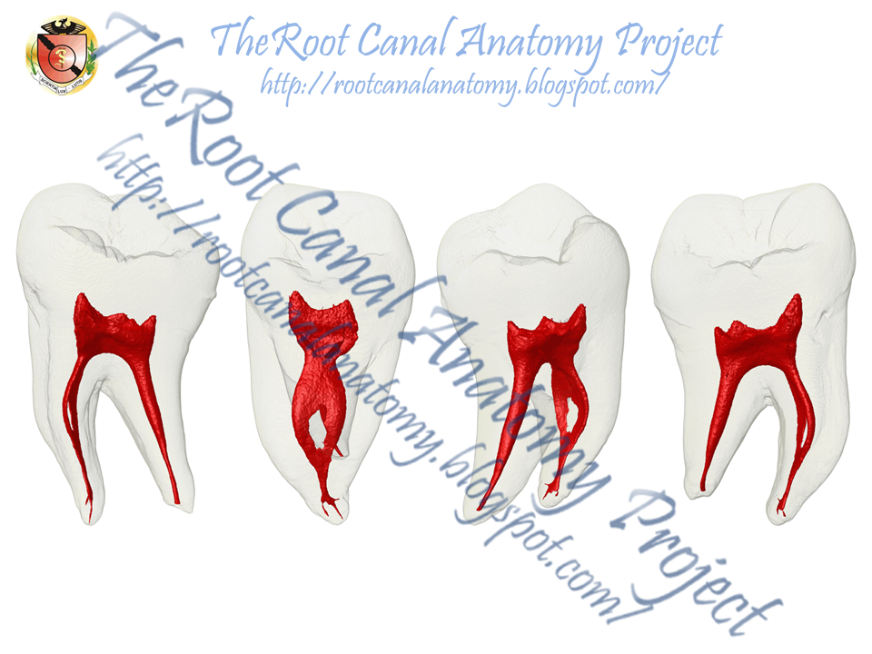
The Root Canal System of Mandibular First Molar
The root canal system of the mandibular first molar is one of the most complex in the human dentition. Typically, the mesial root contains two canals (mesiobuccal and mesiolingual), while the distal root has one canal. However, variations such as the presence of a third canal in the mesial root (mesiobuccal 2 or MB2) are common.
Anatomical Variations in the Root Canal System
Studies have shown that the mandibular first molar exhibits a wide range of anatomical variations, including:
- Additional canals in the mesial root
- Fused or separate roots
- Curved or straight canals
These variations can significantly impact the success of root canal treatment, emphasizing the need for thorough preoperative evaluation.
Read also:Cane Corso With Chain A Comprehensive Guide To Owning And Training This Majestic Breed
Clinical Significance of Root Canal Anatomy
Understanding the root canal anatomy of the mandibular first molar is clinically significant for several reasons:
- Improved treatment outcomes
- Reduced risk of complications
- Enhanced patient comfort
Clinicians who are well-versed in the anatomy of this tooth are better equipped to handle challenging cases and achieve optimal results.
Diagnostic Tools for Root Canal Anatomy
Modern diagnostic tools have revolutionized the way clinicians approach root canal treatment. Some of the most effective tools include:
- Digital radiography
- Cone-beam computed tomography (CBCT)
- Operating microscopes
These technologies enable practitioners to visualize the intricate details of the root canal system, facilitating accurate diagnosis and treatment planning.
Treatment Protocols for Mandibular First Molar
Treating the mandibular first molar requires a systematic approach. Key steps in the treatment protocol include:
- Access cavity preparation
- Canal identification and negotiation
- Canal shaping and cleaning
- Canal obturation
Following these steps meticulously is essential for ensuring the success of endodontic therapy.
Common Complications in Root Canal Treatment
Despite advancements in technology and techniques, complications can still occur during root canal treatment. Common issues include:
- Canal blockages
- Perforations
- Instrument separation
Proper training and experience can help minimize the risk of these complications, ensuring safer and more effective treatments.
Recent Research and Advancements
Research in the field of endodontics continues to evolve, with new studies shedding light on the anatomy of the mandibular first molar. Recent advancements include:
- Improved imaging techniques
- New canal preparation instruments
- Advanced obturation materials
These innovations are paving the way for more precise and efficient treatments, ultimately benefiting both clinicians and patients.
Conclusion and Future Directions
In conclusion, the mandibular first molar root canal anatomy is a complex and fascinating subject that requires thorough understanding and expertise. By mastering the anatomy, variations, and treatment protocols, clinicians can significantly enhance their ability to deliver successful endodontic care. We encourage readers to:
- Explore further resources and research
- Share their experiences and insights
- Engage with the dental community to stay updated on advancements
Thank you for reading this comprehensive guide. We hope it has provided valuable insights into the world of mandibular first molar root canal anatomy. For more articles on dental topics, visit our website and join the conversation.