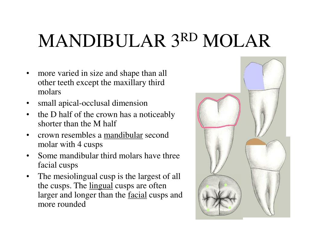
The mandibular first molar roots are one of the most critical components of the human dentition, playing a significant role in mastication and overall oral health. Understanding the structure and function of these roots is essential for dentists, dental students, and anyone interested in oral anatomy. This article will delve into the intricacies of mandibular first molar roots, exploring their anatomy, clinical significance, and related dental procedures.
As a foundational aspect of dental health, mandibular first molars are the first permanent molars to erupt in the mouth, typically around the age of six. These teeth are pivotal in maintaining proper occlusion and supporting facial structure. Their roots provide stability and strength, making them vital for chewing and digestion.
In this comprehensive guide, we will explore the anatomy of mandibular first molar roots, their variations, and the clinical implications associated with them. By the end of this article, you will have a thorough understanding of why these roots are so crucial in dental health and how they impact various dental treatments.
Read also:Exploring Mlwbdlove A Comprehensive Guide To Understanding Benefits And Risks
Table of Contents
- Anatomy of Mandibular First Molar Roots
- Biological Function and Importance
- Variations in Mandibular First Molar Roots
- Clinical Significance of Mandibular First Molar Roots
- Root Canal Treatment for Mandibular First Molars
- Extraction Process of Mandibular First Molars
- Common Disorders Affecting Mandibular First Molar Roots
- Preventive Care for Mandibular First Molars
- Research Insights and Recent Developments
- Conclusion
Anatomy of Mandibular First Molar Roots
The mandibular first molar typically has two roots: a mesial root and a distal root. These roots anchor the tooth firmly into the alveolar bone, providing stability for chewing and other oral functions. The mesial root is generally larger and more prominent than the distal root.
Root Structure
The roots of the mandibular first molar are covered by cementum, a calcified tissue that helps attach the tooth to the periodontal ligament. Inside the root, the pulp chamber houses the nerve and blood supply, which are essential for the tooth's vitality.
Canal Configuration
One of the critical aspects of mandibular first molar roots is their canal configuration. Most mandibular first molars have two canals in the mesial root and one canal in the distal root. However, variations can occur, making root canal treatment more complex.
Biological Function and Importance
The mandibular first molar roots play a crucial role in the biological function of the tooth. They provide structural support, enabling the tooth to withstand the forces of mastication. Additionally, the roots help maintain the integrity of the alveolar bone, preventing bone resorption.
Role in Occlusion
The roots of the mandibular first molars are instrumental in maintaining proper occlusion. They ensure that the teeth align correctly, allowing for efficient chewing and digestion. Proper occlusion also contributes to the overall aesthetics of the face.
Variations in Mandibular First Molar Roots
While most mandibular first molars have two roots, variations can occur. Some individuals may have a single fused root, while others may have three or more roots. These variations can impact dental procedures and must be carefully evaluated by dentists.
Read also:99 A Comprehensive Guide To Understanding Its Significance And Impact
Causes of Root Variations
Root variations can be attributed to genetic factors, environmental influences, or developmental anomalies. Understanding these variations is essential for accurate diagnosis and treatment planning.
Clinical Significance of Mandibular First Molar Roots
The clinical significance of mandibular first molar roots cannot be overstated. Dentists must have a thorough understanding of root anatomy to perform procedures such as root canal treatment, extractions, and implants effectively.
Diagnostic Techniques
Modern diagnostic techniques, such as digital radiography and cone-beam computed tomography (CBCT), allow dentists to visualize the roots of mandibular first molars in great detail. These tools help identify root variations and plan treatment accordingly.
Root Canal Treatment for Mandibular First Molars
Root canal treatment is a common procedure performed on mandibular first molars. This treatment involves removing the infected or damaged pulp tissue from the root canals and filling them with a biocompatible material.
Steps in Root Canal Treatment
- Access the pulp chamber through the crown of the tooth.
- Locate and clean the root canals using specialized instruments.
- Shape the canals to facilitate the filling process.
- Fill the canals with gutta-percha and a sealer.
- Restore the tooth with a crown or filling material.
Extraction Process of Mandibular First Molars
In some cases, the extraction of mandibular first molars may be necessary due to severe decay, trauma, or orthodontic reasons. The extraction process involves carefully removing the tooth, including its roots, from the alveolar bone.
Post-Extraction Care
After extraction, patients must follow proper post-operative care to ensure proper healing. This includes avoiding strenuous activity, maintaining oral hygiene, and taking prescribed medications as directed.
Common Disorders Affecting Mandibular First Molar Roots
Several disorders can affect the roots of mandibular first molars, including root resorption, periodontal disease, and periapical abscesses. Early diagnosis and treatment are crucial to preventing further complications.
Treatment Options
Treatment options for root-related disorders vary depending on the severity of the condition. Conservative treatments, such as scaling and root planing, may be sufficient for mild cases, while surgical intervention may be necessary for more severe conditions.
Preventive Care for Mandibular First Molars
Preventive care is essential for maintaining the health of mandibular first molars and their roots. Regular dental check-ups, proper oral hygiene practices, and a balanced diet can help prevent decay and other dental issues.
Tips for Maintaining Root Health
- Brush twice daily with fluoride toothpaste.
- Floss daily to remove plaque between teeth.
- Visit the dentist regularly for professional cleanings and exams.
- Avoid sugary snacks and beverages that can contribute to decay.
Research Insights and Recent Developments
Recent research has shed light on the complexities of mandibular first molar roots, offering new insights into their anatomy and treatment. Advances in imaging technology and regenerative medicine are paving the way for innovative approaches to dental care.
Future Directions
Future research may focus on developing new materials and techniques for root canal treatment and regenerating damaged root structures. These advancements could revolutionize the way dentists approach mandibular first molar root-related issues.
Conclusion
Mandibular first molar roots are a vital component of the human dentition, providing stability and support for one of the most important teeth in the mouth. Understanding their anatomy, variations, and clinical significance is crucial for dentists and patients alike. By following preventive care guidelines and seeking professional treatment when necessary, individuals can maintain the health of their mandibular first molars and enjoy optimal oral health.
We invite you to share your thoughts and experiences in the comments section below. Additionally, feel free to explore other articles on our site for more information on dental health and related topics. Together, we can promote a healthier, happier smile for everyone!

